Human Generated Data

Title

A Section of the Human Bloodstream. Capillary Vessel

Date

1930, printed late 1950s

People

Artist: Carl Strüwe, German 1898 - 1988

Classification

Photographs

Credit Line

Harvard Art Museums/Busch-Reisinger Museum, Antonia Paepcke DuBrul Fund, 2016.105

Copyright

© Carl Strüwe / Steven Kasher Gallery

Human Generated Data

Title

A Section of the Human Bloodstream. Capillary Vessel

People

Artist: Carl Strüwe, German 1898 - 1988

Date

1930, printed late 1950s

Classification

Photographs

Credit Line

Harvard Art Museums/Busch-Reisinger Museum, Antonia Paepcke DuBrul Fund, 2016.105

Copyright

© Carl Strüwe / Steven Kasher Gallery

Machine Generated Data

Tags

Amazon
created on 2019-04-09

Art 98.5
Drawing 98.5
Doodle 97.5
Rug 89.6
Sketch 74.6
Sea 68.6
Outdoors 68.6
Water 68.6
Nature 68.6
Ocean 68.6
Pattern 61.4
Sea Life 58.9
Reef 58.9
Animal 58.9
Coral Reef 57.4
Graphics 57
Plant 56.9

Clarifai
created on 2018-02-19

art 97.4
illustration 97.2
print 96.3
desktop 96.1
texture 93.8
pattern 93.7
no person 92.6
old 92.5
symbol 92.5
design 92.3
vintage 92
ancient 91.9
one 91.8
people 91.7
retro 89.7
antique 89.5
decoration 88.7
closeup 88
culture 86.9
engraving 85.7

Imagga
created on 2018-02-19

arabesque 35.5
design 31.3
art 30.8
texture 30.6
pattern 30.1
decoration 27.8
floral 27.2
vintage 26.5
retro 26.2
lace 25.8
old 25.8
antique 25.3
paper 25.1
grunge 23
style 20.8
wallpaper 20.7
decor 20.3
fabric 20.2
graphic 19.7
ornate 19.2
ornament 19
flower 18.5
backdrop 18.1
drawing 17.3
mosaic 16.3
decorative 15.9
element 15.7
leaf 14.8
paisley 14
card 13.9
ancient 13.8
traditional 13.3
ornamental 13.3
artistic 13
tile 13
embroidery 12.9
frame 12.5
textile 12.3
textured 12.3
color 12.2
stamp 12.1
effect 11
creative 10.6
plant 10.5
brown 10.3
silhouette 9.9
material 9.8
shape 9.7
detail 9.7
black 9.6
used 9.6
old fashioned 9.5
symbol 9.4
money 9.4
classic 9.3
letter 9.2
stencil 9.1
aged 9.1
postage 8.8
bookmark 8.8
faded 8.8
scrapbook 8.7
mail 8.6
holiday 8.6
line 8.6
finance 8.5
elegance 8.4
damask 8.4
cover 8.3
foliage 8.3
cash 8.2
dirty 8.1
transducer 8
postal 7.9
obsolete 7.7
exchange 7.6
oriental 7.6
swirl 7.4
light 7.4
artwork 7.3
business 7.3
square 7.2
border 7.2
painting 7.2
collection 7.2
wealth 7.2

Google
created on 2018-02-19

black and white 93.4
flora 82.9
monochrome photography 76
monochrome 73.4
organism 72.6
tree 71.7
drawing 65.8
art 64.7
visual arts 60.2
pattern 59.8
plant 59.2
printmaking 57.9
font 56.9
illustration 56.4
artwork 55
graphics 50.7

Microsoft
created on 2018-02-19

text 100
book 99.5

Color Analysis

Feature analysis

Amazon

Rug
Rug 89.6%

Categories

Captions

Microsoft
created by unknown on 2018-02-19

a close up of a book 55.7%
close up of a book 49%
a close up of a book cover 48.9%

Clarifai
created by general-english-image-caption-blip on 2025-05-12

a photograph of a black and white photograph of a plant with leaves -100%

OpenAI GPT

Created by gpt-4o-2024-05-13 on 2024-12-30

The image appears to be a black and white microscopic view of a biological sample, possibly a cross-section of tissue. It shows intricate, branching structures that resemble villi or glandular formations typically found in the lining of the intestine or other tubular organs. The fine and detailed patterns suggest the presence of cellular or sub-cellular elements, with some areas stained darker to highlight specific features or cellular structures within the tissue. The overall appearance is highly textured with a clear distinction between different regions, indicating a high level of complexity.

Created by gpt-4 on 2024-11-20

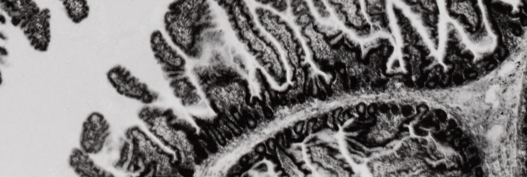
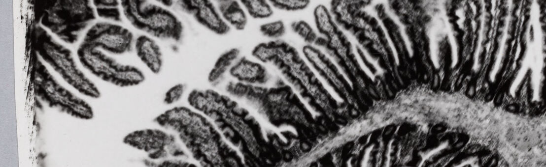
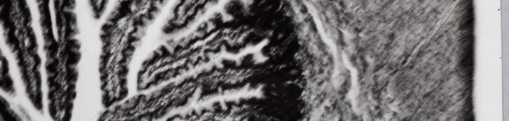
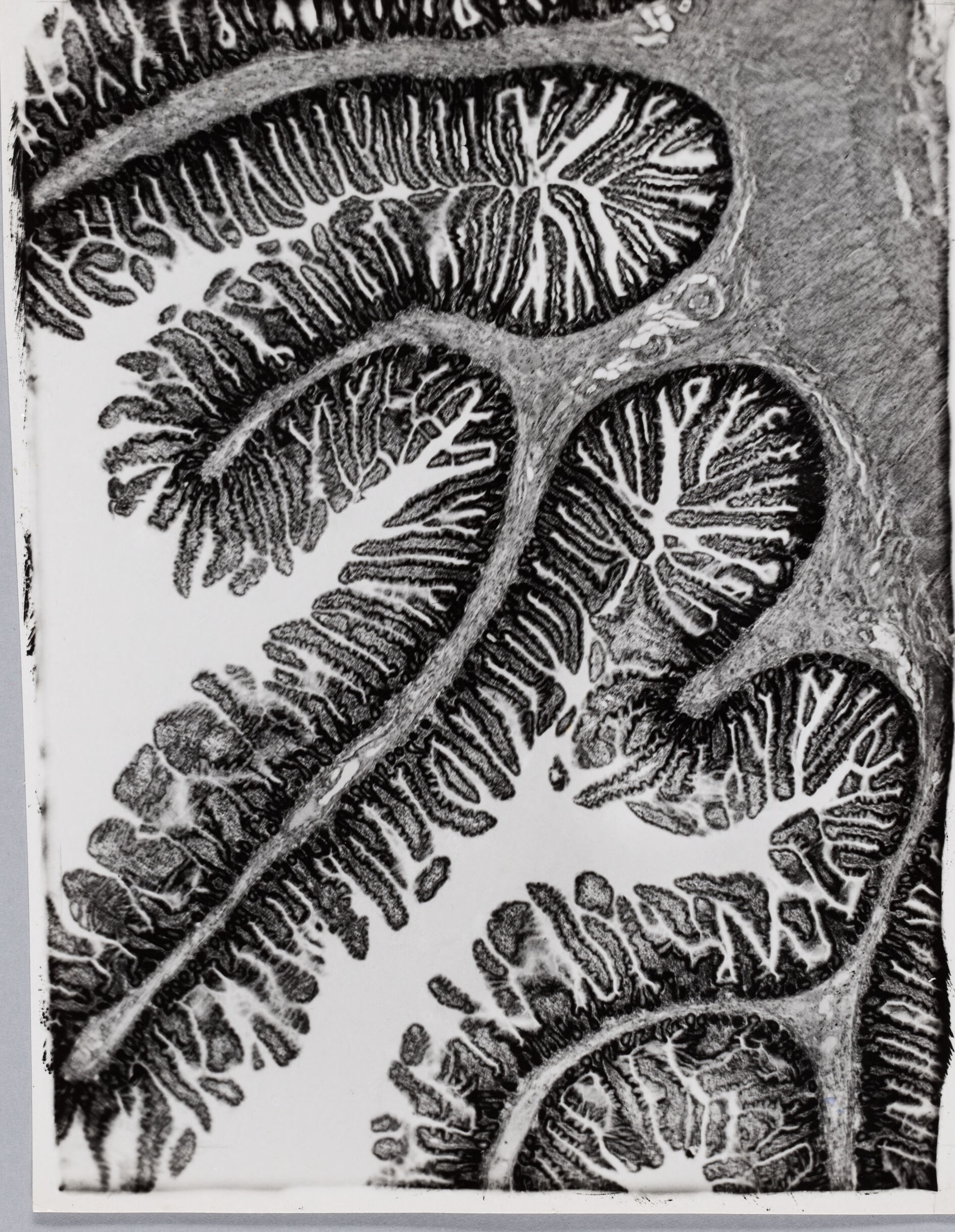
This is a monochrome image of what appears to be a microscopic view of biological structures, likely a section of tissue. The structures resemble the shape and pattern of villi found in the small intestine, where the finger-like projections increase the surface area for absorption of nutrients. Each of the projections has a patterned texture that could indicate the presence of cells or microvilli on the surface. The contrast in the image highlights these textures and creates a clear distinction between the different parts of the tissue. The overall image gives a sense of intricate organic patterns and is representative of the complexity found within biological organisms.

Anthropic Claude

Created by claude-3-5-sonnet-20241022 on 2024-12-27

This appears to be a microscopic image showing cross-sections of intestinal villi. The image is in black and white and shows the characteristic finger-like projections that line the intestinal wall. These villi are crucial for increasing the surface area of the intestine, allowing for better absorption of nutrients from food. The branching, tree-like patterns visible in each villus show the internal structure, including what appears to be the lamina propria and possibly blood vessels. The image has a scientific, almost artistic quality to it, with the stark contrast between the dark tissue structures and the white background creating an intricate, organic pattern.

Created by claude-3-haiku-20240307 on 2024-11-20

The image appears to be a close-up black and white photograph of the cross-section of a citrus fruit, likely an orange or grapefruit. The image captures the intricate and detailed patterns of the fruit's internal structure, revealing the segmented sections and the intricate webbing of the fruit's pulp and rind. The image has a striking textural quality, with the dark and light contrasts creating a dramatic visual effect that highlights the organic, natural beauty of the fruit's anatomy.

Meta Llama

Created by us.meta.llama3-2-90b-instruct-v1:0 on 2024-12-24

This image appears to be a microscopic view of a cross-section of the small intestine, showcasing its intricate structure. The image is in black and white, with the tissue sample stained to highlight its various components.

The small intestine is a long, thin tube that plays a crucial role in the digestion and absorption of nutrients from food. It is lined with finger-like projections called villi, which increase the surface area for absorption. The villi are covered with tiny hair-like structures called microvilli, which further enhance the absorption process.

In this image, the villi are visible as dark, branching structures that protrude from the wall of the intestine. The microvilli are not visible at this magnification, but they would be present on the surface of the villi. The image also shows the muscular layer of the intestine, which is responsible for peristalsis, the movement of food through the digestive tract.

Overall, this image provides a detailed view of the small intestine's structure and function, highlighting its importance in the digestive process.

Created by us.meta.llama3-2-11b-instruct-v1:0 on 2024-12-01

The image appears to be a microscopic view of a section of the small intestine, possibly from a human or animal specimen. The image is in black and white, with the tissue appearing dark gray and the background appearing light gray.

The small intestine is shown in cross-section, with the lumen (the innermost part of the intestine) visible at the top of the image. The lumen is lined with villi, which are small, finger-like projections that increase the surface area for absorption of nutrients.

The villi are surrounded by a layer of epithelial cells, which are the cells that line the inside of the intestine. These cells are responsible for absorbing nutrients from the food that passes through the intestine.

The image also shows the muscularis mucosae, which is a layer of smooth muscle that helps to move food through the intestine. This layer is visible as a thin, dark line that runs along the edge of the villi.

Overall, the image provides a detailed view of the structure of the small intestine, highlighting the different layers of tissue that make up this important organ.

Amazon Nova

Created by amazon.nova-lite-v1:0 on 2025-06-04

The image is a detailed, black-and-white medical illustration depicting a section of human tissue, possibly from the colon or small intestine. The illustration shows a series of circular structures that appear to be villi, which are finger-like projections that increase the surface area for absorption in the intestines. The villi are intricately detailed, with a complex network of blood vessels and lymphatic vessels visible within them. The overall texture and pattern resemble a leaf or fern, with a branching structure that extends from the base of each villus. The image has a highly magnified and detailed appearance, suggesting it was created using a microscope or other specialized imaging technique.

Created by amazon.nova-pro-v1:0 on 2025-06-04

The image is a black-and-white illustration of a microscopic view of what appears to be a biological structure, possibly from a plant or an organism. The structure consists of elongated, branching elements that resemble the vasculature or circulatory system of a plant, with intricate patterns of veins or vessels. The details are highly magnified, showcasing the complexity and intricacy of the biological system. The image is likely a scientific illustration or a photograph taken under a microscope, aimed at studying the anatomy and physiology of the organism.

Google Gemini

Created by gemini-2.0-flash on 2025-05-24

The image appears to be a black and white photograph, possibly a close-up or microscopic view, of biological tissue. The structure shows several elongated, finger-like projections or folds that curve and twist, resembling intestinal villi. The interior of these projections has a branching pattern reminiscent of tree branches or veins. There is a contrast between the dark outer layer and the lighter inner structure, which helps to define the shapes and patterns. The image has a texture that suggests it may have been produced through a technique like etching or a printing process that emphasizes detail and contrast.

Created by gemini-2.0-flash-lite on 2025-05-24

The image is a black and white microscopic view of a tissue sample. It appears to be a cross-section of the intestinal lining, showcasing the characteristic villi (finger-like projections) and crypts (depressions) of the intestinal mucosa.

Here's a breakdown:

  • Villi: The numerous, elongated, and branching structures represent the villi. These structures are responsible for increasing the surface area of the intestine, aiding in nutrient absorption. They have a dark, almost striated appearance.
  • Crypts: The spaces between and surrounding the villi.
  • Color/Tone: The image's grayscale palette accentuates the tissue structures and the texture of the sample.
  • Overall Impression: This is a detailed, artistic depiction of the intestinal structure, which likely comes from a biological or medical context.

Mistral Pixtral

Created by us.mistral.pixtral-large-2502-v1:0 on 2025-06-03

The image appears to be a microscopic view of a section of tissue, likely from the gastrointestinal tract, such as the small intestine. The tissue shows characteristic features of intestinal villi and crypts. Here are some detailed observations:

  1. Villi: The finger-like projections extending into the lumen (the open space) are villi. These structures increase the surface area for absorption of nutrients in the intestine.

  2. Crypts of Lieberkühn: The invaginations or depressions between the villi are known as crypts of Lieberkühn. These are the sites where intestinal stem cells reside and where cell proliferation and differentiation occur.

  3. Epithelial Lining: The villi and crypts are lined with epithelial cells. These cells are responsible for the absorption of nutrients and the secretion of digestive enzymes.

  4. Lamina Propria: The tissue underlying the epithelial lining is the lamina propria, which contains connective tissue, blood vessels, and immune cells.

  5. Muscularis Mucosae: Beneath the lamina propria, there is a thin layer of smooth muscle known as the muscularis mucosae, which helps with the movement and folding of the mucosal layer.

  6. Staining: The image is likely stained to enhance the contrast between different tissue components. The darker areas could represent nuclei or other cellular structures, while the lighter areas might represent extracellular matrix or cytoplasm.

This detailed structure is essential for the digestive and absorptive functions of the intestine.

Text analysis

Amazon

eUNIulK
AENRM
eUNIulK SWEAL
CARSSACE
SWEAL