
Machine Generated Data
Tags
Color Analysis
Feature analysis
Amazon
Clarifai

AWS Rekognition
| Adult | 94.4% | |


Categories
Imagga
created on 2018-10-05
| pets animals | 86.1% | |
| paintings art | 6.4% | |
| food drinks | 5.3% | |
| people portraits | 1.6% | |
| interior objects | 0.3% | |
| macro flowers | 0.1% | |
| text visuals | 0.1% | |
Captions
Microsoft
created by unknown on 2018-10-05
| a man sitting in front of a computer | 42.6% | |
| a man sitting in front of a computer screen | 35.9% | |
| a man looking at a computer screen | 35.8% | |
Clarifai
No captions written
Salesforce
Created by general-english-image-caption-blip on 2025-05-26
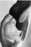
a photograph of a person holding a piece of food in their hand
Created by general-english-image-caption-blip-2 on 2025-07-10
a film strip with two pictures of a dog
OpenAI GPT
Created by gpt-4o-2024-08-06 on 2025-06-17
The image is a vertical strip resembling a sequence of frames from a film reel or film strip. The film strip contains six black and white frames stacked vertically. Each frame shows a close-up of a small animal, likely a tiny bird or chick, being held gently by a person’s fingers. The focus is on the chick, showing different angles and details in each frame. The film strip features the classic perforated edges commonly seen in photographic or cinematic film. The background is blurred, emphasizing the animal and the hand. The lighting is consistent across the frames, showing a soft texture on the chick and the fingers holding it.
Created by gpt-4o-2024-11-20 on 2025-06-17
The image depicts a vertical filmstrip containing several sequential frames of a close-up view of what appears to be a seed emerging from or interacting with a textured surface. The seed-like object is held or manipulated by fingers in each frame, showing a gradual progression or movement. The black-and-white photography highlights the detailed textures of the object and its surroundings, emphasizing the tactile and organic qualities. Each frame displays slight changes in the position or state of the object, suggesting growth, extraction, or examination.
Anthropic Claude
Created by anthropic.claude-3-haiku-20240307-v1:0 on 2025-06-17
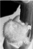
The image appears to be a series of black and white photographs depicting a small, furry animal, likely a kitten or young cat. The photographs show the animal in various poses, curled up and resting. The images capture the soft, fluffy texture of the animal's fur and its delicate features. The overall impression is one of gentleness and serenity.
Created by us.anthropic.claude-3-5-sonnet-20241022-v2:0 on 2025-06-17
This appears to be a series of black and white x-ray or radiographic images showing a tooth or dental structure from slightly different angles. The images are stacked vertically and show what looks like a molar or premolar tooth, with clear visibility of the tooth's internal structure, root, and crown. The contrast in the images helps highlight the different densities within the tooth, which is useful for dental diagnosis and treatment planning.
Meta Llama
Created by us.meta.llama3-2-90b-instruct-v1:0 on 2025-06-02
The image depicts a strip of film featuring a sequence of six black-and-white frames. Each frame showcases a close-up view of a person's face, with their mouth open and a white, fuzzy object visible inside. The object appears to be a piece of food, possibly a marshmallow or a similar soft treat.
The person's face is partially obscured by shadows, making it difficult to discern any distinct features. The background of the image is dark, which further emphasizes the focus on the person's mouth and the object within.
The overall atmosphere of the image suggests that it may be a still from a film or video that captures a moment of eating or consuming something. The use of black-and-white tones adds a sense of nostalgia and timelessness to the image, while the close-up perspective creates an intimate and engaging visual experience.
Created by us.meta.llama3-2-11b-instruct-v1:0 on 2025-06-02
The image is a black and white film strip featuring a close-up of a person's face, with their head tilted back and mouth open. The person has short hair and appears to be wearing a dark shirt or jacket. The film strip consists of six frames, each capturing a slightly different angle of the person's face. The background of the image is dark and indistinct, but it appears to be a studio setting with a plain backdrop.
The overall effect of the image is one of drama and intensity, as the person's open mouth and tilted head create a sense of tension and emotion. The use of black and white film adds to the dramatic effect, creating a sense of timelessness and classic Hollywood glamour.
Amazon Nova
Created by amazon.nova-lite-v1:0 on 2025-06-04
The image is a black-and-white sequence of photographs showing a series of frames from a medical or scientific study. Each frame appears to capture a close-up view of a biological specimen, possibly a part of an animal or a plant, undergoing a process. The specimen in the center of each frame is somewhat obscured by a shadow or a dark area, suggesting it might be inside a container or under a microscope. The background is uniformly dark, enhancing the contrast and detail of the specimen. The sequence shows the specimen in different stages, possibly indicating a progression of time or a series of events. The overall impression is of a detailed, high-contrast study of a small object.
Created by amazon.nova-pro-v1:0 on 2025-06-04
The image appears to be a series of black and white photographs, likely taken in a laboratory setting. The images show a close-up view of what appears to be a biological specimen, possibly a piece of tissue or an organ. The specimen has a distinct, irregular shape with a textured surface. The images are arranged in a sequence, suggesting a time-lapse or sequential documentation of the specimen's condition or changes over time. The overall composition and presentation of the image suggest a scientific or medical context, possibly related to research or analysis of biological samples.
Google Gemini
Created by gemini-2.0-flash on 2025-05-28
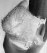
The image shows a strip of photographic film in black and white. The film contains several frames, each capturing a similar scene. The scene appears to be a close-up of what seems to be an animal or object with fur or a textured surface. It is partially obscured or being touched by a darker object or figure, which could be a hand or part of another object. The frames are stacked vertically, creating a sequence of slightly varying perspectives or moments of the same subject. The overall tone of the image is stark due to the high contrast and monochrome palette.
Created by gemini-2.0-flash-lite on 2025-05-28
Here's a description of the image:
The image is a strip of 35mm film with six sequential frames of a close-up shot, presented in black and white, with the light areas seeming very bright. Each frame seems to be focused on a fluffy object.
The fluffy object seems to be held by a hand or fingers, and partially obscured by a dark, possibly cylindrical, object. The frames show slight variations in the position of the hand and the fluffy object.
Mistral Pixtral
Created by us.mistral.pixtral-large-2502-v1:0 on 2025-06-08
The image appears to be a series of six X-ray or CT scan slices showing a lateral view of a knee joint. Here is a detailed description of what is visible in each slice from top to bottom:
Top Slice:
- The topmost image shows the upper part of the knee joint.
- The femur (thigh bone) and the patella (kneecap) are visible.
- The joint space between the femur and the patella is clearly seen.
Second Slice:
- This slice shows a slightly lower section of the knee joint.
- The femur and the patella are still visible.
- The joint space is more pronounced, and the initial part of the tibia (shin bone) begins to appear.
Third Slice:
- This image shows a more central part of the knee joint.
- The femur, patella, and tibia are all visible.
- The joint space between the femur and tibia is more defined.
Fourth Slice:
- This slice shows a lower section of the knee joint.
- The femur, patella, and tibia are visible.
- The joint space is clearly seen, and the fibula (the smaller bone in the lower leg) may start to appear.
Fifth Slice:
- This image shows an even lower section of the knee joint.
- The femur, patella, tibia, and fibula are visible.
- The joint space is still visible, but the bones are more prominent.
Bottom Slice:
- The bottommost image shows the lowest section of the knee joint.
- The femur, patella, tibia, and fibula are visible.
- The joint space is less pronounced, and the bones are more clearly defined.
Overall, the series of images provides a comprehensive view of the knee joint from different angles, showing the interaction between the femur, patella, tibia, and fibula. This type of imaging is typically used to diagnose and assess conditions affecting the knee joint, such as fractures, dislocations, or degenerative diseases like arthritis.
Qwen
Created by Qwen/Qwen2.5-VL-7B-Instruct on 2025-07-19
The image is a sequence of six black and white photos arranged vertically, resembling a filmstrip. Each photo shows a close-up of a small, furry animal, likely a young hedgehog or a similar creature, being held gently by a person's hand. The animal appears to be very small and is nestled in the person's palm, with its head and front limbs visible. The hand is positioned to support the animal securely. The lighting is soft, and the background is out of focus, drawing attention to the animal and the hand. The sequence seems to capture a gentle and tender moment of interaction between the person and the small animal.
Created by Qwen/Qwen2.5-VL-72B-Instruct on 2025-07-19
This image is a black-and-white photographic sequence showing the process of cleaning an animal's teeth. The sequence is presented in six frames, arranged vertically.
- The first frame shows a close-up of an animal's mouth, likely a dog or a cat, with its teeth and gums visible. A person is holding the animal's mouth open.
- The second frame shows the introduction of a dental tool, possibly a scaler, being used to clean the teeth.
- The third frame continues to show the dental tool being used, with the person carefully maneuvering it around the teeth.
- The fourth frame shows the continuation of the cleaning process, with the dental tool still in use.
- The fifth frame shows the dental tool being removed from the animal's mouth.
- The sixth frame shows the animal's mouth after the cleaning process, with the person holding the mouth open to inspect the teeth.
The sequence effectively demonstrates the step-by-step process of dental cleaning for an animal.