Machine Generated Data
Tags
Color Analysis
Feature analysis
Amazon
| Fish | 90.7% | |
Categories
Imagga
| paintings art | 99.7% | |
Captions
Microsoft
created on 2019-08-10
| a close up of text on a white surface | 36.9% | |
| a close up of text on a white background | 36.8% | |
| close up of text on a white surface | 33.3% | |
OpenAI GPT
Created by gpt-4o-2024-05-13 on 2025-02-11
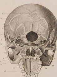
The image is a detailed anatomical illustration of the human skull and its parts. It includes various views and sections of the skull, such as lateral (side), superior (top), frontal, and internal views. The illustration appears to be labeled with letters and numbers to identify different parts of the skull, which is often seen in medical or anatomical illustrations for educational purposes. The details show an emphasis on the bone structure and the intricate connections between different parts of the skull.
Created by gpt-4 on 2025-02-11
Anthropic Claude
Created by claude-3-5-sonnet-20241022 on 2025-02-11
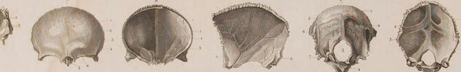
This is a detailed anatomical illustration plate showing various views and sections of human skulls. The top row features three lateral (side) views of skulls. Below that are several smaller illustrations showing different cross-sections and detailed views of skull bones and cranial anatomy. The bottom row shows three larger views including: a sectioned skull showing internal anatomy, a frontal view of the skull base, and a front view of a complete skull. The illustrations appear to be done in a classical medical illustration style with fine shading and careful attention to anatomical detail. This type of illustration would typically be found in medical or anatomical textbooks from the 18th or 19th century. The image has a designation "B.8" in the top right corner, suggesting it's part of a larger series of anatomical plates.
Created by claude-3-opus-20240229 on 2025-02-11
The image shows an anatomical illustration of human skulls from different angles. The top row depicts three views of the skull in profile, showing the cranium, jawbone, and various bony landmarks. Below that are additional detailed views focusing on specific regions of the skull, such as the facial bones, base of the skull, and individual bones that make up the cranium. The illustrations have labels indicating different parts of the skull anatomy. The drawing style appears to be from an older medical or anatomical textbook, using shading to provide a three-dimensional appearance to the skulls and bones.
Created by claude-3-haiku-20240307 on 2025-02-11
This image appears to be a scientific illustration or diagram depicting various views and cross-sections of human skulls. The main focus is on the overall structure and anatomy of the skull, showing different perspectives and levels of detail. The image does not contain any identifiable human faces or individuals, but rather presents the skulls in an objective, scholarly manner for the purposes of study and analysis.
Meta Llama
Created by us.meta.llama3-2-11b-instruct-v1:0 on 2025-03-18
The image is a detailed illustration of the human skull, showcasing various views and sections. The illustration is rendered in a monochromatic color scheme, with a light beige background and darker shading used to highlight the different features of the skull. **Key Features:** * **Multiple Views:** The illustration presents multiple views of the skull, including front, side, and rear perspectives. * **Sectional Views:** Additionally, the illustration includes sectional views of the skull, providing a detailed look at the internal structure. * **Anatomical Details:** The illustration highlights various anatomical details, such as the shape and size of the cranium, the position and arrangement of the facial bones, and the structure of the teeth and jaw. * **Labels and Numbers:** The illustration includes labels and numbers that identify specific parts of the skull, allowing for easy reference and understanding. * **Artistic Style:** The illustration is rendered in a realistic style, with attention to detail and accuracy in depicting the anatomy of the human skull. **Overall Impression:** The illustration provides a comprehensive and detailed view of the human skull, making it an excellent resource for students, medical professionals, and anyone interested in anatomy. The use of multiple views and sectional cuts allows for a thorough understanding of the skull's structure and function.
Created by us.meta.llama3-2-90b-instruct-v1:0 on 2025-03-18
The image is a vintage illustration of human skulls, showcasing various views and sections. The illustration features four skulls in different orientations, with the top row displaying three skulls from the side, front, and back, respectively. The bottom row presents three skulls with their tops removed, revealing the interior structure. The illustration also includes several smaller images of skull fragments, such as the nasal cavity, eye sockets, and jawbone, which provide a detailed view of the skull's anatomy. The background of the illustration is a light beige color, with a subtle texture that resembles aged paper. The overall effect is one of a classic anatomical drawing, likely created for educational or scientific purposes.
Amazon Nova
Created by amazon.nova-pro-v1:0 on 2025-02-11
The image is a black-and-white illustration of skulls and their various parts. The illustration is arranged in a grid-like format, with three rows and four columns. The top row features three skulls, each with a different orientation. The second row contains six smaller illustrations of the skulls' parts, such as the jawbone, eye socket, and nasal cavity. The bottom row features three more skulls, each with a different orientation.
Created by amazon.nova-lite-v1:0 on 2025-02-11
The image is a scientific illustration depicting various views and sections of a human skull. It includes detailed drawings of the skull from different angles, such as frontal, lateral, and top views, as well as cross-sections that show the internal structure. The illustrations are in black and white, emphasizing the anatomical features. Some of the drawings highlight specific bones and regions, such as the cranial base, facial bones, and the interior of the skull, providing a comprehensive view of the skull's anatomy.
Text analysis
Amazon