Machine Generated Data
Tags
Color Analysis
Face analysis
Amazon

AWS Rekognition
Age | 43-51 |
Gender | Female, 61.3% |
Calm | 99.9% |
Sad | 0.1% |
Happy | 0% |
Confused | 0% |
Angry | 0% |
Disgusted | 0% |
Surprised | 0% |
Fear | 0% |
Feature analysis
Categories
Imagga
paintings art | 99% | |
Captions
Microsoft
created by unknown on 2022-01-23
diagram | 68.5% | |
Clarifai
created by general-english-image-caption-blip on 2025-05-22
a photograph of a man standing in front of a machine | -100% | |
Meta Llama
Created by us.meta.llama3-2-11b-instruct-v1:0 on 2025-05-24
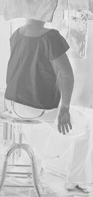
The image depicts a medical setting, likely from the early 20th century, featuring a patient undergoing a radiographic examination. The patient is positioned on a chair, wearing a lead apron and a lead mask covering their head and neck. A technician stands beside them, dressed in a long coat and holding a control panel.
The room is equipped with various medical equipment, including a large X-ray machine and a fluoroscope. The walls are made of brick, and the floor appears to be tiled. The overall atmosphere suggests a clinical environment where medical professionals are conducting a diagnostic procedure using radiation technology.
The image provides a glimpse into the history of medical imaging and the evolution of radiography techniques. It highlights the importance of radiation safety measures, such as lead aprons and masks, which were introduced to protect patients and staff from harmful radiation exposure.
In summary, the image captures a moment in time when medical technology was still in its early stages of development, and the use of radiation was a relatively new concept. The scene is a testament to the dedication and expertise of medical professionals who worked tirelessly to improve patient care and advance the field of medicine.
Created by us.meta.llama3-2-90b-instruct-v1:0 on 2025-05-24
The image is a black-and-white photograph of a medical setting, likely from the early 20th century. The scene depicts a patient undergoing an X-ray examination, with a medical professional present to assist.
Key Elements:
- Patient: The patient is seated on a chair, facing away from the camera. They are wearing a dark-colored shirt and light-colored pants. A large, rectangular device covers their head and upper body, which appears to be an early X-ray machine.
- Medical Professional: A medical professional, possibly a doctor or radiologist, stands beside the patient. They are dressed in a long coat and tie, indicating their professional attire. The professional's face is blurred, making it difficult to discern any facial features.
- X-ray Machine: The X-ray machine is a prominent feature in the image. It consists of a large, rectangular box with a circular opening at the top. The machine is positioned in front of the patient, with the circular opening aligned with the patient's head and upper body.
- Background: The background of the image shows a brick wall with windows, suggesting that the medical facility is located in a building with a traditional architectural style. The overall atmosphere of the image is one of a clinical setting, with a focus on the medical procedure being performed.
Historical Context:
The image likely dates back to the early 20th century, when X-ray technology was still in its infancy. The use of X-rays for medical imaging was first introduced in the late 19th century, but it wasn't until the early 20th century that X-ray machines became more widely available and commonly used in medical settings. The image provides a glimpse into the early days of medical imaging and the development of X-ray technology.
Conclusion:
The image is a fascinating glimpse into the history of medical imaging and the development of X-ray technology. It highlights the importance of medical innovation and the role of medical professionals in advancing our understanding of the human body. The image also serves as a reminder of the significant progress that has been made in medical imaging over the past century, and the continued importance of investing in medical research and innovation.
Amazon Nova
Created by amazon.nova-lite-v1:0 on 2025-05-23
The black-and-white image shows a person sitting on a stool in a room with a large machine. The person is wearing a mask and appears to be undergoing a procedure. A woman in a lab coat is standing behind the person, holding a device. The room has brick walls and a tiled floor. There are several machines and equipment in the room, including a large machine with a wheel and a metal structure.
Created by amazon.nova-pro-v1:0 on 2025-05-23
The image is in black and white. It features a person sitting on a stool in a room with a brick wall. The person is wearing a black top and pants. The person's head is covered with a cloth. The person is looking to the right. There is a person standing in front of the person, wearing a jacket and pants. The person is holding a wire and a wheel. There are several machines in the room.
Google Gemini
Created by gemini-2.0-flash-lite on 2025-05-18
This appears to be a photograph of an early X-ray examination. Here's a breakdown of what I see:
- Setting: The image is of an interior room, likely a doctor's office or hospital room. The background includes windows and brick walls.
- Equipment: There is a large X-ray machine prominently in the frame, with a tube to emit the X-rays, a stand, and other related equipment. The room also seems to be filled with different panels.
- Subjects: Two people are visible. One, who is seated, appears to be the patient. They are wearing a dark top and are likely having their head examined, judging by the apparatus in front of them. The other person is likely a doctor or technician, standing beside the machine and holding a wheel and what looks like a cord.
- Overall Impression: The image portrays a very early stage of medical technology in the 20th century. The equipment appears to be less refined than modern counterparts, and there isn't much in the way of radiation protection.
Created by gemini-2.0-flash on 2025-05-18
Here is a description of the image:
The image shows a black and white photograph of what appears to be an early X-ray examination setting. A person, presumably the patient, is seated on a stool with their back to the viewer, possibly positioned for a scan. The patient has a cover or shield placed over their head.
A person dressed in a long coat is standing to the side, holding a large wheel, perhaps operating the machine. The room is filled with equipment, including a large, bulbous object on a stand, possibly the X-ray source, and other box-like apparatus with meters and dials. The scene is very bright, characteristic of a film negative. The overall setting gives the impression of a historic medical procedure.
Mistral Pixtral
Created by us.mistral.pixtral-large-2502-v1:0 on 2025-05-23
The image depicts an early X-ray procedure being performed in what appears to be a medical or laboratory setting. Here are the key details:
Patient and Equipment:
- A patient is seated on a stool, facing away from the camera, with their arm extended. The patient appears to be wearing a protective garment or apron, likely to shield them from radiation.
- There is an X-ray machine positioned in front of the patient. The machine has a large, adjustable frame with a circular component that likely houses the X-ray tube.
Operator:
- A person, presumably a medical professional or technician, stands to the right of the patient. This individual is also wearing protective clothing, which suggests an awareness of the need for radiation safety.
- The operator appears to be adjusting or monitoring the equipment, possibly preparing to take an X-ray image.
Setting:
- The room contains various pieces of equipment and machinery, indicating a specialized environment, likely a radiology department or laboratory.
- There are multiple pieces of equipment visible, including what appears to be a control panel or generator on the right side of the image.
Historical Context:
- The overall appearance of the equipment and clothing suggests that this image is from the early to mid-20th century, a period when X-ray technology was still relatively new and evolving.
This image provides a glimpse into the early days of radiology, showcasing the equipment and procedures used at the time.